Top videos

This is Part 3 of a 3 part series video, where I will be talking about wisdom tooth pain, what to expect when you visit the dentist for wisdom tooth pain, and what to do after extraction of your problematic wisdom tooth!
If you want to know more about what Wisdom teeth actually are and why they cause trouble, then check out my first video of this series here
https://www.youtube.com/watch?v=u-dIE...
The best way to treat your Wisdom Tooth pain is not always surgery. There are many options available that can help to treat the symptoms while still saving your tooth from removal!
So, if you're wondering about what to do if you are experiencing Wisdom Tooth Pain and what are the various treatment options then check out the Episode 2 of this series here
https://www.youtube.com/watch?v=FfFkFinY7Dk
What do I use to shoot my videos?
I use a basic ring light which I purchased from Amazon. It helps me shoot in low light conditions and comes with a decent tripod stand Link - https://amzn.to/2Z7p2mP
For audio I use Amputive Collar Mic which can be purchased by clicking on this link - https://amzn.to/3e7oGAE
For the camera, I just use my OnePlus 6. I use iMovie to edit my videos.
Follow me on social media :
Instagram : https://www.instagram.com/doctor.upasana
Facebook : https://www.facebook.com/ismile.india/
Website : https://www.i-smile.in/
Google Page : https://g.page/iSmile

Wisdom teeth are also called third molars and it is not unusual for these teeth to become "stuck", wedged in or what we call impacted. They are the last teeth to come through into the mouth and often there is not enough room for them. Most people have four wisdom teeth but up to 10% of the population missing one or more. When wisdom teeth become impacted they cause a number of problems.
![[ENG] Extraction of horizontal Lt. Mn third molar](https://i.ytimg.com/vi/uYZeObkoTkk/maxresdefault.jpg)
Title : Extraction of horizontal Lt. Mn third molar
Case by Dr. Cho Yongseok
👉 [ Master of Third molar extraction ] by Dr. Cho YongSeok
Full lectures are available on the Dental Bean site.
View lecture : https://www.dentalbean.com/product/productDetail.aspx?_cateKey=&_prKey=2087&_lMenuKey=1005
How to take more lectures?
We invite you to our global web site www.dentalbean.com
Sign up on our global site, enjoy all of great doctor’s special lectures!
Dental bean is open learning place for dentists all over the world.
Dental clinical study to learn in anywhere & anytime.
https://www.dentalbean.com
#Dentalbean #DentalEducation #Online #Dentist #Dentistry #Dental #Diente #Implant
#Implantology #Education #Lecture #Onlineclass #Webinar #Seminar #Sinus #Bonegraft
#GBR #Vitalroot #Thirdmolar
In this video, Dr. H. Ryan Kazemi, Oral and Maxillofacial Surgeon in Bethesda, MD, describes how maxillary sinus perforation may occur following teeth extractions and demonstrates a 3-layer technique for its management.

In this video, you will learn helpful tips on what to do to prevent bleeding, what to do to stop bleeding after tooth extraction, and how to troubleshoot in times of prolonged bleeding.
You will also learn of what your dentist should be focusing on prior dental extraction, preventative measurements that may need to be taken, and what to do in case you are bleeding more than expected.
Please do not forget to like the video and subscribe to the channel!
https://deluxedentistry.com - to learn more or to become our patient.

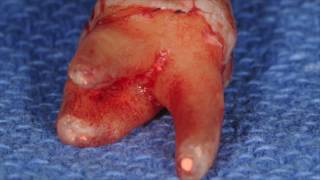
how to remove root tip, which was left after tooth extraction. Implant placed , area bone grafted.
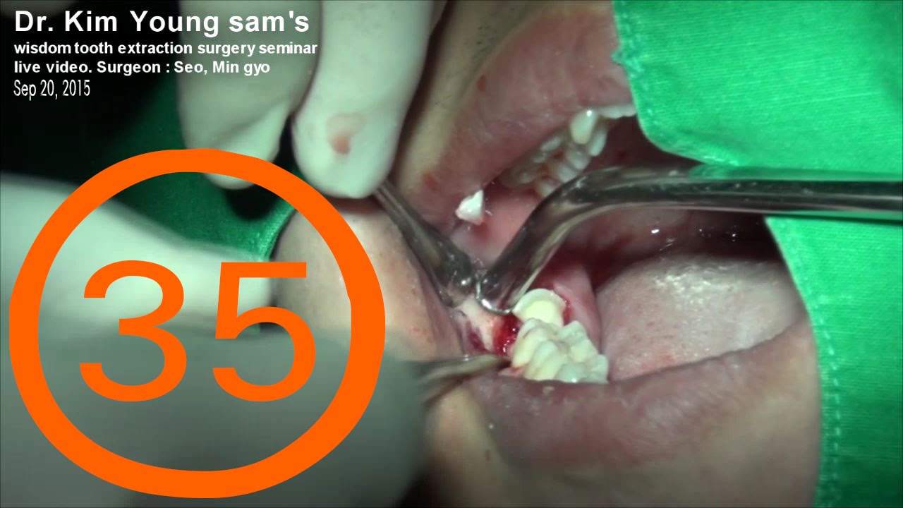
Dr. Kim Young sam's wisdom tooth extraction surgery seminar
live video on Sep 20 in 2015. Surgeon : Seo, Min gyo
2015년 9월 20일 김영삼원장의 사랑니 발치 세미나의 라이브 서저리
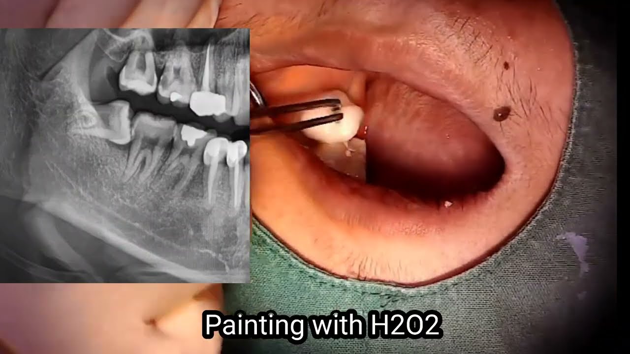
Surgical extraction of mandibular third molar(Lower wisdom tooth) With radiographic description.
Warnings: Graphic Content of Surgery. Viewer discretion advised.
Wisdom Tooth Removal in Amarillo TX: Kristen | Oral & Maxillofacial Surgery of the Panhandle Region - http://panhandleoralsurgery.com
Wisdom teeth, or Third molars, are an additional set of teeth, which grow under the surface of the gums and, in many cases, are undetectable without proper x-ray evaluation or 3-D scanning. It is not uncommon for teens and adults to be unaware that these wisdom teeth exist in their mouth until they begin to present significant problems. In some cases, the third molars may fully grow into the mouth or partially grow into the mouth and additional issues may arise. Unfortunately, the majority of people do not have a long enough jaw bone or a large enough mouth to accommodate these teeth. Wisdom tooth removal by a licensed Oral and Maxillofacial surgeon will provide the highest quality surgical expertise for the surgical process.
As an oral and maxillofacial surgeon, Dr. Bryan Bailey at Oral and Maxillofacial Surgery of the Panhandle Region manages a variety of issues relating to the mouth, gums, teeth, and general facial region. He practices a full scope of oral and maxillofacial surgery with expertise in wisdom teeth removal, tooth/teeth extractions, various dental implant procedures, corrective jaw surgery (orthognathic surgery), oral pathology, and bone grafting. We provide surgery services to Amarillo, TX, and surrounding Panhandle region of Texas. He is an expert in numerous aspects of facial surgery including facial trauma and reconstructive surgery, dental implants, bone grafting, and orthognathic surgery.
Hi, my name is Kristen. I live in Amarillo. I came to Dr. Bailey to get my wisdom teeth taken out. It was an all right experience. I don’t really remember anything, but the staff treated me great. It was not as bad as I expected it to be. If you live in Amarillo and you need to get your wisdom teeth taken out, come to Dr. Bailey because he’s great!
Oral and Maxillofacial Surgeryof the Panhandle Region
4905 Lexington Square
Amarillo, TX 79119
Ph: 806.367.9990
Fax: 806.367.9945
Hours:
Mon, Tu & Thur: 8:30a - 5:00p
Wed: 8:30a - 1:00p
Fri: 8:30a - 12:00p
Our Services:
Wisdom Teeth | Dental Implants | Impacted Tooth | Tooth Extraction | Bone Grafting | Oral Pathology | Facial Trauma | CT Scans | Expose & Bonds
Cities Served:
Amarillo, TX | Panhandle Region, TX | Potter County, TX | Randall County, TX | Canyon, TX | Hereford, TX | Clarendon, TX | Pampa, TX | Dumas, TX | Borge, TX | Fritch, TX | Stinnett, TX | Spearman, TX | Stratford, TX | Dalhart, TX | Friona, TX | Tulia, TX | Plainview, TX | Floyada, TX | Childress, TX | Tucumcari, NM

At Alexandria Oral Surgery & Dental Implant Center, we will do everything possible to save a natural tooth. If extraction is the best option for you, your surgeon can create a treatment plan to remove your teeth efficiently and safely at one of our offices in Alexandria, LA, or Vidalia, LA. Sometimes tooth decay, injuries, and other problems lead to irreparable tooth damage. When restorative treatments, such as root canals, can’t save a tooth, removal may be the only option to prevent infections from spreading. Our surgeons can also provide options to replace your missing teeth, such as dental implants.
Dr. Smith received his undergraduate degree in Biochemistry at Occidental College in 1988, after which he was part of a research team at Children’s Hospital of LA studying retinal cancer. He then decided to continue his education by earning his law degree at the University of Oregon. As a lawyer, he fiercely advocated for the right of his clients, and he carried that passion for helping people over into his medical career. He earned his Doctor of Medicine from Oregon Health & Sciences University, followed by a general surgery internship at Swedish Medical Center in Seattle. He was drawn to the precise techniques involved in oral surgery, and completed an oral surgery residency program at the University of Louisville in 2006.
Alexandria Oral Surgery & Dental Implant Center
https:/alexandriaos.com
Fred W. Smith, DMD, MD, JD
P. Steven Arnold Jr., DMD, MD
Alexandria
1403 Peterman Dr
Alexandria, LA 71301
Natchitoches
140 E 5th Street
Natchitoches, LA 71457
Services Offered:
Dental Implants | Wisdom Teeth Removal | Tooth Extractions | Bone Grafting | Oral Pathology Treatment | Dental Implant-Supported Dentures | Pre-Prosthetic Surgery | Treatment of Sleep Apnea | Exposure of Impacted Canines | Apicoectomy | Stem Cell Banking | Facial Cosmetics
Cities Served:
Alexandria | Pineville | Natchitoches | Marksville | Winnfield | Derioder | Mansura | Bunkle | Boyce | Manny | Leesville | Opelousas | Oakdale | Vidalia
Have You Been Wanting To Improve Your Confidence In Oral Surgery And Extract More Teeth? Do You Want An Experienced Dentist That You Can Contact Anytime To Support Your Efforts? Join Me. The O.S. Accelerator Course: https://onlineexodontia.com/store
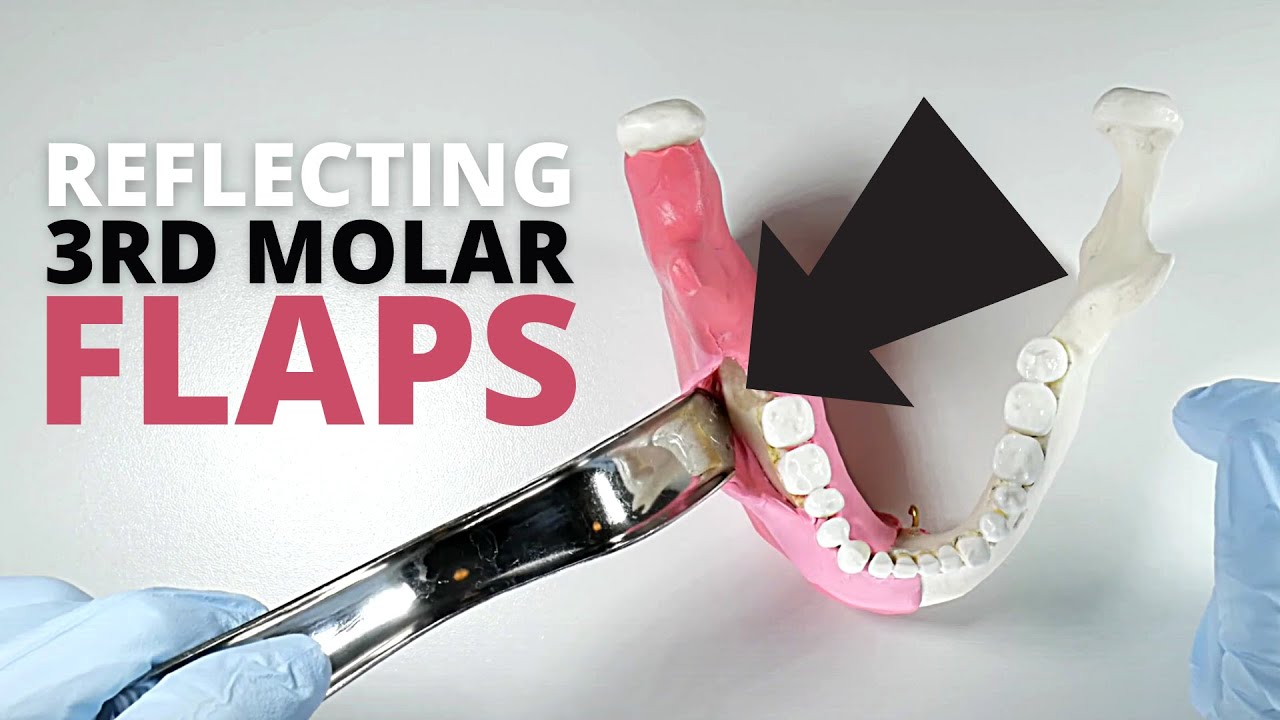
This video discusses key tips for simplified reflection of full-thickness mucoperiosteal flaps in the third molar region. A look at common retractors, incision techniques, help tips are covered in the video. Maintaining a clean, accessible, well-lit surgical field is key to doing proper surgery.

Apicoectomy is used to eliminate an infection in the root of a tooth and the surrounding tissues. Here you will find all the information about this treatment.
00:00 Introduction:
3D video of Clínica Médico Dental Pardiñas (http://j.mp/cPardinas) about apicoectomy, a dental treatment to remove an infection that affects the root apex of a tooth and adjacent tissues.
00:10 What is apicoectomy?
The apicoectomy is a procedure to remove an infection that affects the root of a tooth and the adjacent tissues. It is performed in cases where the endodontic treatment has failed, presence of canals or false canals unsealed or in teeth where the access to the apex is not possible by a conventional root canal treatment.
00:40 How is an apicoectomy performed?
To do so, an incision is performed, and sufficient bone is removed to get access to the root apex. Then the infectious cyst is removed cleaning and irrigating the area. A beveled shape cut is made to the root removing the apex and sealing the root with different materials. The area is sutured and will regenerate until its complete healing.
More info about apicoectomy 👉 http://bit.ly/apicoectomyCP
Subscribe to our Youtube channel 👉 http://bit.ly/suscribeteCP
MORE 3D VIDEOS in English 👉 http://j.mp/dentistry3D
Follow us also on:
👍 Instagram: https://www.instagram.com/clinicapardinas
👍 Facebook: https://www.facebook.com/clinicapardinas
👍 Twitter: https://twitter.com/ClinicaPardinas
👍 Linkedin: https://www.linkedin.com/company/clinica-pardinas
👍 Tiktok: https://www.tiktok.com/@clinicapardinas/
👍 Pinterest: https://www.pinterest.es/clinicapardinas/
#apicoectomy #oralsurgery #dentalabscess

Dentistry by Dr. Tim Kosinski. Atraumatic extraction of lower molar #18 utilizing the Physics Forceps lower universal forcep from Golden Dental Solutions (Dr. Richard Golden), the inventors and manufacturers of the Physics Forceps. Depending on access, posterior physics forceps may have to be used on 2nd and 3rd molars. In this case, the standard series lower universal Physics Forceps was used since the tooth was accessible. Note, we recommend to use the bumper guards on the forcep, which was not used in this case.
Please visit www.physicsforceps.com or www.goldendentalsolutions.com to learn more about and/or to purchase the Physics Forceps.
Address:
Golden Dental Solutions
27115 Gratiot Avenue,
Suite B Roseville,
Michigan 48066.
Telephone: 1.877.987.2284
E-mail: info@goldendentalsolutions.com

This video demonstrates technique for surgical removal of impacted wisdom tooth under local anaesthetic
website: tt-os.co.uk
removal of the tooth with bone attached to it , ankylosis .When removing tooth, peace of bone came out with root.

Hi i am Dr Rahul Narkhede
Dentist at smile care 32 dental clinic n implant centre , in kamothe ,navi mumbai since more than 9 yrs.
About this video:
Wisdom teeth are the last teeth to erupt from mouth and most of the time we need to extract them bcoz of limited space and inability to maintain oral hygiene.
Brushing technique video link...
https://youtu.be/SfoYmEPQxbA
Flossing technique video link....
https://youtu.be/jmn39y6qh8U
To book appointment with me...
www.smilecarekamothe.com

#Learningbasalimplants
Heyyyy 😃
Welcome to the Yorkshire Dental Suite Youtube channel. We are a Dental Super clinic based in England, Leeds and absolutely love what we do. This year we have started our journey to shaping the future of dentistry in the UK and would like to bring you with us on that journey 😊 Subscribe for weekly uploads and feel free to follow us on Instagram @yorkshiredentalsuite https://www.instagram.com/york....shiredentalsuite/?hl
Feel free to comment below any video suggestions for us to create as we want to give you guys the content you want !
For more information on our treatments/what we do visit our website www.yorkshiredentalsuite.co.uk or email us hello@yorkshiredentalsuite.co.uk
Enjoy the video and we hope to see our Youtube family soon at the clinic !! 😃
High Quality Surgical videos and uncut stories ▶ https://surgeoncut.com
Wisdom tooth removal or third molar removal is a run off the mill type surgery performed at Richardsons dental and Craniofacial Hospital, TamilNadu, India.
What were the patients complaints?
This patient in his fourth decade of life, had recurrent food impactions in the region and that also caused him a lot of pain.
This was occurring very often. As often as once in a couple of months.
What was done at Richardsons?
A cone beam CT scan was taken and the position of the tooth was clearly studied in relation to the inferior alveolar nerve. The tooth was lying down in a forward position, called mesioangular.
The surgery of dis impaction was planned under local anesthesia, however in the operation theatre.
How long did it take?
With the experienced team at Richardsons dental and Craniofacial hospital, it hardly took 20 minutes to retrieve the tooth.
Later on, as you can clearly see in the video, sutures were placed with vicryl 3.0. The advantages of using this material is that the sutures don’t have to be removed.
When would the sutures be removed?
They need not be removed as resorb-able stitches were used.
How long is the healing?
Healing not only depends on the type of the impacted wisdom tooth, but it also depends
On how efficiently and effectively the surgeon does the job of delivering the tooth.
Usually doesn’t take more than a few days.
Where is Richardsons?
Located in Nagercoil, Tamil Nadu, India
Social media :
Instagram: drsunilrichardson
Twitter: facesurgeon1
What’s App : +919443182860
Email : drsunilrichardson@hotmail.com
Websites :
www.facesurgeon.in
www.drsunilrichardson.com
LinkedIn: drsunilrichardson

Surgical extraction of vertically impacted 48 by Dr Nauv Kashyap. Demonstration video for dentists.
www.raceviewdental.com.au