


![[Hue Dental] Impacted Wisdom Tooth Extraction Wisdom Teeth Removal Procedure Oral Surgery](https://i.ytimg.com/vi/oGpZss0ikEM/maxresdefault.jpg)


Removal of a molar tooth root fragment in a horse - tooth root abscess part 2
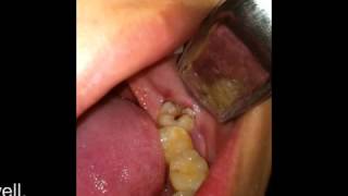
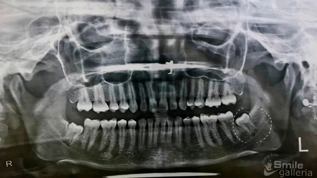
This horse was featured on a previous video. The tooth had fractured secondary to dental decay. The tooth had fragmented and had been partially removed. Instead of subjecting the horse to a general anaesthetic, which carries a small but significant risk, it was sedated using a morphine drip. The alveolar socket was anaesthetised by placing a maxillary nerve block and a gingival block. The tooth root fragment & alveolar socket was examined using an oroscope. This was also used to facilitate sectioning of the tooth root using an 3mm bone cutting bur. This was only partially successful as only several small fragments were removed. The decision was made to repulse the remaining tooth root fragment. Local anaesthetic was infiltrated under the skin in a position over the tooth root. A Coomer bone drill was used to drill a hole in the caudal maxillary sinus. A blunt ended trocar pin was placed over the tooth root and a mallet was used to drive the fragment through the alveolar socket. Radiograph were taken of the molar arcade revealing two further fragments (cementomas) located in the alveolar socket. These were relatively easily removed under oroscopic guidance. The resultant wound and sinus was flushed using sterile saline.The skin wound was closed with staples. The surgery had left hole (fistula) between the oral cavity (mouth) and sinus. This was packed with dental putty to prevent a sinusitis occurring due to food contamination from the mouth. These wounds can take up to 6 months to heal and need repacking regularly with dental putty.